

dalszy na tkaninie, zdjęcie dalszy na tkaninie, tkanina dalszy
FILTRUJ WYNIKI
Orientacja obrazów: wszystkie
pozioma
pionowa
kwadratowa
Rodzaj obrazów: wszystkie
zdjęcie
ilustracja
wektor
Zastosuj
#771498628
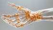 3D imaging of the human hand bones and joints, hightech medical detail, clipart isolated on a white...
#504132321
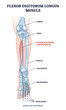 Flexor digitorum longus muscle with human leg and foot bones outline diagram. Labeled educational...
#535878261
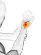
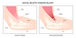 distal bicep tendonitis rotator cuff pain upper arm inflamed
#549423259
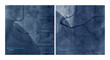
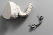 Dental casting gypsum model of human jaws. Crooked teeth and distal bite. Shots were made before...
#533921152
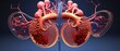
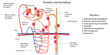 Diuretics and the kidney. Vector illustration
#478777958
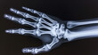
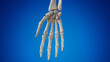 3d rendered illustration of the hand bones
#308477432
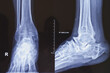
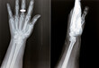
 Film X ray wrist radiograph show distal forearm bone broken ( distal end radius fracture). The...
#250700669
 Dental casting gypsum model of human jaws. Crooked teeth and distal bite. Shots were made before...
#651516367
 A Man with torn biceps does preacher curls. Working out and injury rehabilitation after 6 months of...
#693861601
 Male, 68 years old, chest pain for 7 hours. Coronary angiography suggests occlusion of the distal...
#780312014
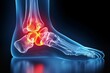
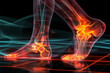 Illustration of human foot with ankle pain. anatomy, physical therapy concept
#766518609
 Depict the salivary glands and ducts within the oral cavity using X-ray illustration.
#702293903
 Film xray x-ray or radiograph of a hand and fingers showing the numbers one through five 1-5. One,...
#167788616
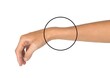
 man on the beach with a bandage in his wrist
#786399098
 A 3D model of the human skeleton, highlighting the spinal column for orthopedic educational purposes
#755653879
 X-ray of ankle joint both view. Diffuse sclerosis at tarsal, metatarsal bones and distal shaft of...
#652769872
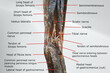 anatomy of popliteal region with the distal portion of back of thigh and proximal portion of back of...
#621988855
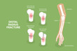 3D Isometric Flat Vector Conceptual Illustration of Distal Radius Fracture, Labeled Educational...
#488023849
 Wisdom Tooth Impaction Infographics
#771444841
 Painful foot, 3d rendered medical illustration
#693362572
 Colles' fracture of an old female, a type of fracture of the distal forearm in which the broken end...
#528102183
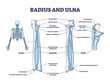 Radius and ulna bone anatomy with arm skeletal structure outline diagram. Labeled educational scheme...
#558260314
 Avascular necrosis hand displacement break painful radial dislocated fall onto an outstretched...
#748972160
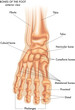 Medical illustration of the main parts of the bones of the foot in anterior view, with annotations.
#475617662
 A photo of plain radiograph on dark background in hospital. The film use for diagnosis the illness...
#535718344
 3d rendered medically accurate illustration of a man having a painful wrist
#308477436
 Film X-ray wrist radiograph show lower end of forearm bone broken (distal end radius fracture) from...
#536599038
 Colles Fracture is a complete fracture of the radius bone close to wrist. Broken wrist.
#698749191
 Human adult hand bones x-ray image. Medical and anatomy radiography or imagery
#587263363
 Goldilocks Buttercup (Ranunculus auricomus). Cauline Leaf Closeup
#780312900
 Illustration of human foot with ankle pain. anatomy, physical therapy concept
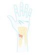
#97973273
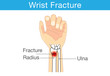 Diagram of wrist have bone fracture
dodaj do ulubionych