

Obraz chrząstka na płótnie, płótno chrząstka, chrząstka canvas
FILTRUJ WYNIKI
Orientacja obrazów: wszystkie
pozioma
pionowa
kwadratowa
Rodzaj obrazów: wszystkie
zdjęcie
ilustracja
wektor
Zastosuj
#731467777
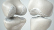
 care bones and knee joints, vitamins, minerals, calcium, zinc, and magnesium. absorbed into...
#698899097
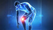
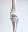
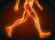
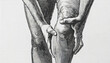
 The leg and knee bone showing pain. medical use Education and Commerce
#473549316
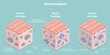
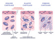 Perichondrium as hyaline, fibrous and elastic cartilage membrane outline diagram. Labeled...
#677519794
 3D Visualization Maps the Comprehensive Anatomy of the Human Knee
#663062890
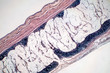
 Showing Light micrograph Type of Tissue Human under the microscope in Lab.
#749956669
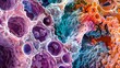 colorful Mesenchyme of structure under the electron microscope.
#193424835
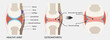
 Knee joint with cartilage loss due to Arthose, front and back- 3D Rendering
#719064494
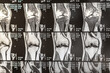
 X-ray shot of a thigh tendon injury
#457697436
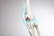
 Knee cartilage bone and muscles pain, human leg anatomy illustration
#729715069
 Painful shoulder joints. Frozen shoulder, impingement. 3d illustration
#703952729
 Male doctor and male patient discussing knee joint model It is likely that the focus will be on the...
#498926926
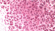 Light microscopy of hyaline cartilage composed of chondrocytes embedded in the cartilage matrix...
#740385958
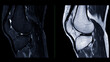 Magnetic resonance imaging or MRI of knee joint. This diagnostic technique is crucial for assessing...
#310224247
 Human hyaline cartilage bone under microscope view for education pathology.
#704559274
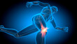 Running man with pain in knee joint - 3D illustration
#686973556
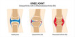 Knee Joint Osteoarthritis vs Rheumatoid arthritis
#694233567
 Rheumatoid arthritis, osteoarthritis of the joint. Images of healthy and diseased joints with main...
#694430488
 human male body gastrocnemius muscles anatomy system. illustration
#740734595
 Close-up of female doctor examining knee joint in clinic. Medical healthcare and orthopedics concept...
#596353798
 3d rendered medical illustration of a wireframe style knee joints
#731309142
 care bones and knee joints, vitamins, minerals, calcium, zinc, and magnesium. absorbed into...
#457928484
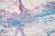
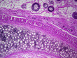 Histology image of elastic cartilage of the epiglottis (100x)
#689718704
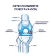 Osteochondritis dissecans or OCD bone and cartilage condition outline diagram. Labeled educational...
#677516484
 3D Visualization Maps the Comprehensive Anatomy of the Human Knee
#620827167
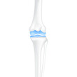 Leg bones knee and joint cartilage healthy. Human skeleton anatomy. Medical health care science...
#553054188
 3D Isometric Flat Vector Conceptual Illustration of Perichondrium, Types of Cartilage
#193328095
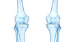
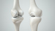 Knee joint with healthy cartilage, front and back- 3D Rendering
#772409611
 tissue engineering and regenerative medicine of skin and cartilage tissue
#460676326
 Knee cartilage bone and muscles pain, human leg anatomy illustration
#539900339
 A surrealistic articular cartilage illustration incorporating modern connection technology. medical...
#583259965
 Joint pain, arthritis and tendon problems. knees with inflammation and pain. Knee and cartilage...
#676629671
 Knee joint x-ray or MRI. Doctor pointed on area of knee joint, where pathology or problem is...
dodaj do ulubionych